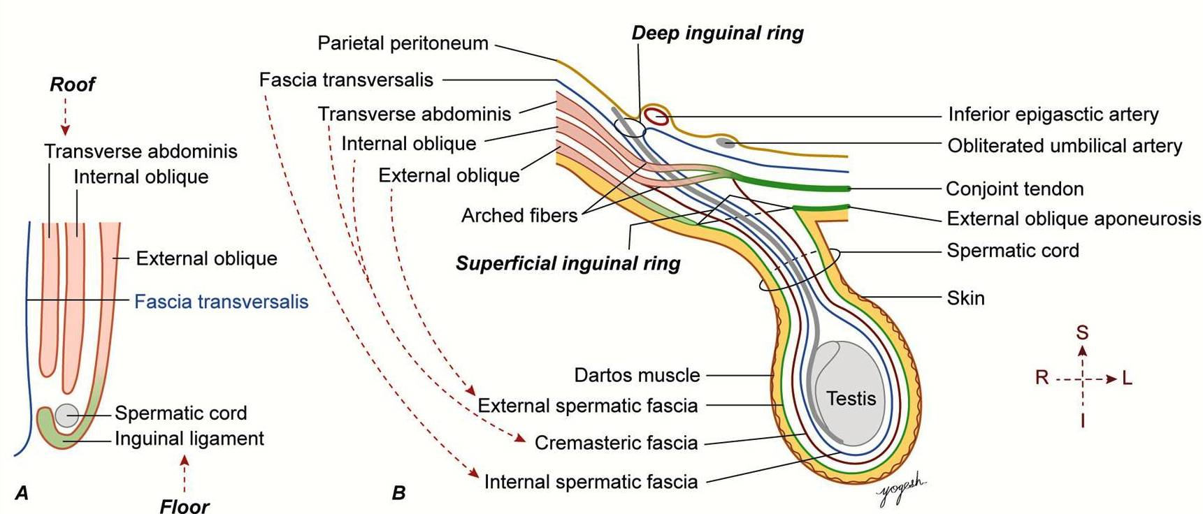
32. Inguinal Canal -Boundaries
Anterior Wall (A): Aponeurosis of the external oblique muscle (along its entire length) and the lateral third of the internal oblique muscle. Posterior Wall (T): Transversalis fascia throughout, reinforced medially by the conjoint tendon (fused internal oblique and transversus abdominis aponeuroses). Roof/Superior Wall (M): Arched fibers of the internal oblique and transversus abdominis muscles. Floor/Inferior Wall (L): Inguinal ligament (a "rolled" edge of the external oblique aponeurosis) and the lacunar ligament medially.